Understanding Pain
Sciatica, Piriformis Syndrome, or a Herniated Disc? How They Differ
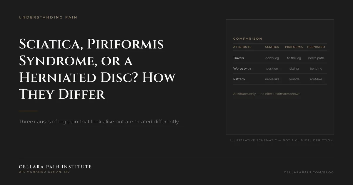
# Sciatica, Piriformis Syndrome, or a Herniated Disc? How They Differ Short answer: "Sciatica" describes where the pain goes, not what causes it. The two most common explanations are a herniated disc irritating a nerve root and piriformis syndrome, in which the sciatic nerve is compressed by a muscle deep in the buttock. They can feel nearly identical from the outside, and they are treated differently — which is why the exam, and sometimes a diagnostic injection, matters more than the label. Nucleus pulposus herniation is the most common cause of sciatic pain and one of the most common indications for spine surgery worldwide, with an estimated prevalence of roughly 1% to 3%.[1] Piriformis syndrome is a clinical diagnosis — sciatic nerve entrapment at the level of the ischial tuberosity — and it is one of the classic mimics that must be separated from a disc problem.[2]
01
Start with the anatomy, because it explains the symptoms
The sciatic nerve is formed by nerve roots that exit the lower spine, then travel through the buttock and down the leg. Two different things can irritate it along that path:
- At the spine: a herniated disc compresses or chemically irritates a nerve root where it exits. This is a radicular problem. Symptoms usually follow a nerve-root map — pain below the knee, into a specific part of the foot, sometimes with numbness in a dermatomal pattern and weakness in a specific muscle group.
- In the buttock: the nerve passes under or through the piriformis muscle. Compression there produces gluteal symptoms — deep buttock pain radiating down the back of the thigh — often provoked by sitting, prolonged standing, or internal rotation of the hip.
Because both conditions send pain down the same highway, location alone is not enough. What differs is the pattern, the triggers, and the exam findings.
02
Five features that help separate them
| Feature | Herniated disc (radicular) | Piriformis syndrome |
|---|---|---|
| Pain location | Follows a nerve root, often below the knee into the foot | Deep buttock, radiating down the back of the thigh |
| Typical trigger | Bending forward, sitting, coughing or sneezing, straight-leg raise | Prolonged sitting, hip rotation, pressure on the buttock |
| Reflexes | May be reduced at the involved level | Typically normal |
| Strength | May show a focal weakness (for example, difficulty with heel or toe walking) | Usually normal, though pain limits effort |
| Response to spine exam | Positive nerve-tension signs | Tenderness over the buttock, pain with resisted hip abduction |
None of these findings is perfect in isolation. The piriformis literature notes that symptoms overlap substantially with other causes of gluteal and radiating leg pain, and that the differential must be worked through deliberately rather than assumed.[2]
03
Why the distinction changes treatment
- Herniated disc / radicular pain: most cases improve over a few weeks with conservative care. Imaging is not indicated without red flags unless symptoms persist beyond roughly six weeks, and MRI is the modality of choice when imaging is needed.[3] When pain persists despite conservative care, targeted options such as epidural steroid injections are considered for short-term relief of radicular pain, and surgery is reserved for selected cases — trials show faster relief with early surgery, but similar outcomes at one to two years.[2][3][4]
- Piriformis syndrome: management typically focuses on activity modification and a graded stretching and strengthening program, with injections or other interventions considered in persistent cases.[2]
- If both are present: this is common and awkward. A person can have a disc bulge on imaging and a tight, irritated piriformis. Treating the wrong one produces the familiar story of a procedure that "didn't work."
That last point explains a lot of patient frustration. An MRI finding is not automatically the pain generator — a point that guidelines reinforce with their repeated caution against treating imaging alone.[5]
04
What the evaluation actually looks like
A focused evaluation should include:
- A pain map — where it starts, where it travels, how far down it goes, and whether it crosses the knee.
- A neurologic exam — reflexes, sensation, and strength by nerve root, plus nerve-tension testing.
- Trigger testing — what makes it worse: bending, sitting, hip rotation, walking.
- Review of existing imaging — with attention to whether the finding matches the symptoms.
- A working hypothesis, stated out loud — for example, "I think this is L5 radicular pain," followed by how it will be tested.
- Selective testing when needed — targeted injections can confirm a nerve root or a muscle as the source when the picture is ambiguous.
05
Red flags that change everything
Seek emergency care for saddle-area numbness, new difficulty starting urination or loss of bowel control, rapidly progressive leg weakness, or fever with new back pain. Cauda equina syndrome is a surgical emergency — roughly 3% of disc herniations — and no elective pathway applies.[6]
For neck and arm symptoms, weakness in both arms or legs, or difficulty with fine hand movements, needs prompt evaluation rather than watchful waiting.
06
FAQ
Can I have sciatica without a herniated disc? Yes. "Sciatica" is a symptom pattern. Disc herniation is the most common cause, but muscle entrapment, spinal stenosis, and other conditions can produce similar leg pain.[1][2]
Does a normal MRI mean it's piriformis syndrome? Not by itself. A normal scan makes a structural disc cause less likely, but piriformis syndrome is diagnosed clinically, and other generators can produce the same pain.[2]
Will stretching fix it? Stretching is part of the plan for piriformis-related pain, but it is rarely sufficient alone, and aggressive stretching during an acute disc flare can worsen nerve pain. The plan should follow the diagnosis.
Do I need an EMG? Electrodiagnostic testing can help distinguish nerve-root involvement from muscle-based pain in ambiguous cases. Whether it is needed depends on the exam findings.
How long before I should be evaluated? If leg pain is not improving after four to six weeks, imaging may become indicated and a specialist evaluation is reasonable.[3]
07
What to do next
The single most useful thing you can bring to an evaluation is the pattern: exactly where the pain starts, how far it travels, and what reliably provokes it. At Cellara Pain Institute in Langhorne, PA, the evaluation is built to answer one question first — is this a nerve root, a joint, or a muscle? — before any treatment is proposed.
Book a consultation to get the pattern read carefully rather than assumed.
08
References
- StatPearls (NCBI Bookshelf). Nucleus Pulposus Herniation. https://www.ncbi.nlm.nih.gov/books/NBK542307/ — accessed 2026-09-16.
- StatPearls (NCBI Bookshelf). Piriformis Syndrome. https://www.ncbi.nlm.nih.gov/books/NBK448172/ — accessed 2026-09-16.
- StatPearls (NCBI Bookshelf). Disk Herniation. https://www.ncbi.nlm.nih.gov/books/NBK441822/ — accessed 2026-09-16.
- American Family Physician. Chronic Low Back Pain in Adults: Evaluation and Management (2024). https://www.aafp.org/afp/2024/0300/chronic-low-back-pain — accessed 2026-09-16.
- American Family Physician. Mechanical Low Back Pain (2018), imaging recommendations. https://www.aafp.org/afp/2018/1001/p421 — accessed 2026-09-16.
- StatPearls (NCBI Bookshelf). Cauda Equina and Conus Medullaris Syndromes. https://www.ncbi.nlm.nih.gov/books/NBK537200/ — accessed 2026-09-16.
Ready to talk about your care?
Book a consultation
A consultation gives you time to discuss your history, questions, and treatment options with a specialist.
Book a Consultation