Understanding Pain
Do You Need an MRI for Back Pain? How Clinicians Decide
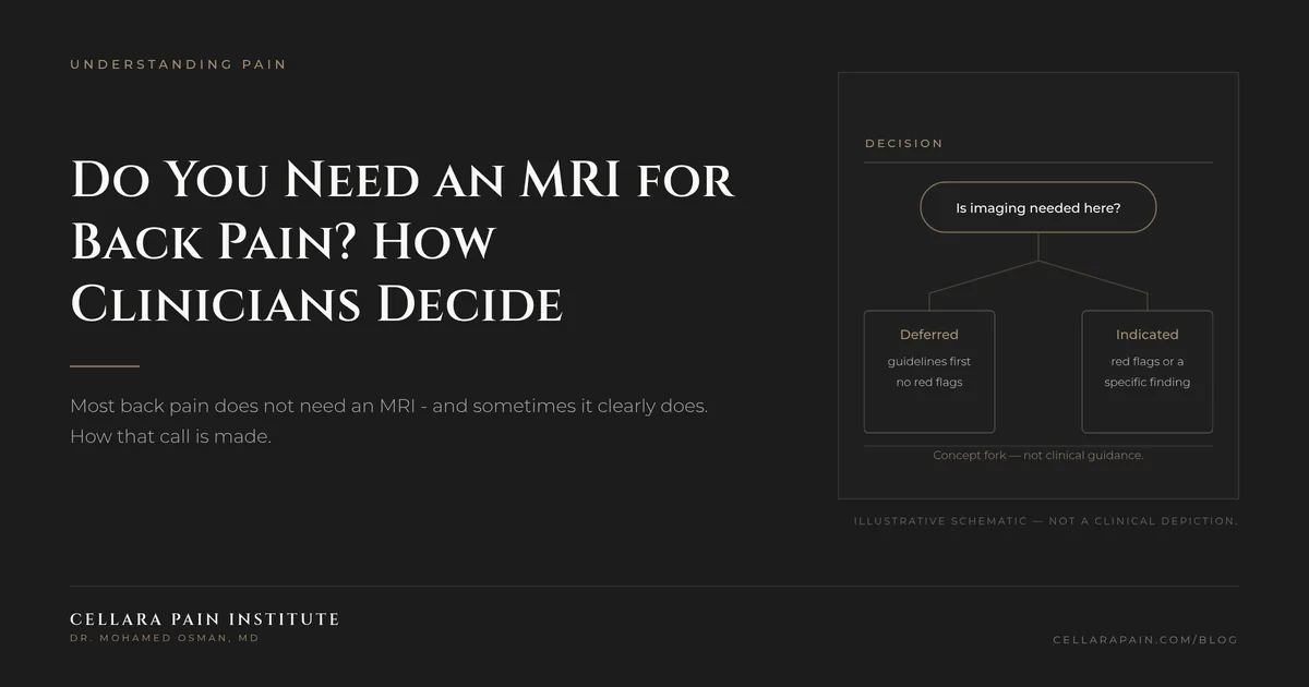
# Do You Need an MRI for Back Pain? How Clinicians Decide Short answer: Usually not at the start — and often yes later. Guidelines consistently recommend against routine imaging for new low back pain without red flags, because most scans find things that are common in people without pain and do not change treatment. Imaging becomes clearly appropriate when there is a red flag, a neurologic deficit, or pain that has not improved with conservative care — commonly at around six weeks. This is one of the few areas where the right answer is genuinely counterintuitive. Patients often expect that a scan will finally explain their pain. Sometimes it does. More often it returns a list of age-related findings that are real but not the pain generator, which can send treatment in the wrong direction.
01
What the guidelines actually say
- Do not order initial imaging studies unless there is concern for cauda equina syndrome, malignancy, fracture, or infection.[1]
- Do not perform imaging for low back pain in the first six weeks unless red flags are present. Red flags include, but are not limited to, severe or progressive neurologic deficits or suspected serious underlying conditions such as osteomyelitis.[1]
- Do not recommend advanced imaging such as MRI within the first six weeks for nonspecific acute low back pain in the absence of red flags.[1]
- Routine imaging is not recommended for chronic low back pain either, but is indicated when red flags are present, when there is a neuromuscular deficit, or when pain does not resolve with conservative therapy.[2]
- When imaging is needed, MRI is the modality of choice.[3]
The pattern is consistent across organizations: imaging is a decision that follows from the clinical question, not a routine step that precedes it.
02
Why "scan everyone" backfires
Imaging findings are common in people who have no pain at all. Disc bulges, disc degeneration, and facet arthritis appear frequently on scans of asymptomatic adults — which means a finding on your report is not automatically your diagnosis.[1][2]
That has two practical consequences:
- A finding can mislead treatment. If a scan shows a disc bulge but the exam points to a facet joint, treating the bulge may produce a procedure that does not help and a conclusion that "nothing works."
- It can create harm without benefit. An unnecessary scan can trigger anxiety, additional testing, and sometimes interventions with their own risks — the classic cascade that the Choosing Wisely recommendations were designed to interrupt.[4]
This is not an argument against imaging. It is an argument for imaging when the result will change what is done.
03
When imaging clearly is appropriate
Bring these to a clinician rather than deciding alone. Imaging is routinely justified when:
- Red flags are present: new saddle numbness, bowel or bladder changes, progressive weakness, fever, unexplained weight loss, night pain that wakes you, cancer history, significant trauma, or long-term steroid use.[1][2]
- There is a neurologic deficit on examination — a focal weakness, a sensory loss in a nerve distribution, or a reflex change.
- Pain has not resolved after a reasonable course of conservative care — commonly around six weeks, and the American College of Radiology appropriateness criteria endorse imaging when there is no improvement after six weeks of conservative medical and physical therapy.[1]
- A procedure is being planned that depends on anatomy — for example, targeting a specific nerve root or planning an intervention.
- Surgery is being considered. A surgical decision should rest on anatomy plus symptoms, which is why guidelines direct imaging to candidates rather than to everyone.[3]
04
Which study, and why MRI is not always the answer
- MRI (without contrast, usually) shows discs, nerve roots, the spinal canal, and soft tissue. It is the modality of choice for most suspected radicular or spinal cord problems.[3]
- X-ray shows alignment, fractures, and advanced degeneration, but not discs or nerves. It is generally not the first study for uncomplicated pain.
- CT is used when bone detail matters or when MRI is not possible.
- Dynamic or weight-bearing imaging is used in specific situations where position changes the picture.
Your exam findings should determine which study is ordered. A scan chosen before the clinical question is defined tends to generate more questions than answers.
05
What belongs in the conversation with your clinician
Ask three questions:
- What are we looking for? A specific suspicion — a nerve root compression, an infection, a fracture — makes the scan interpretable.
- Will the result change my treatment? If not, it may be reasonable to wait and reassess.
- What will we do if the scan is normal? This question keeps the plan moving rather than stalling on a normal result.
06
Red flags that bypass the discussion entirely
Emergency care is the right response to new saddle-area numbness, new difficulty starting urination or loss of bowel control, rapidly progressive leg weakness, fever or chills with new back pain, or severe back pain after trauma. Cauda equina syndrome occurs in roughly 3% of disc herniations and is treated as a surgical emergency.[5]
07
FAQ
Can I just pay for an MRI myself? In many places, yes — but a scan without a clinical question can create confusion and expense. If you self-refer, bring the images to a clinician who will examine you and interpret them against your symptoms.
My MRI shows a herniated disc. Do I need surgery? No. Most disc herniations improve over a few weeks with conservative care, and imaging is not indicated without red flags unless symptoms persist for about six weeks.[3]
How long should I wait before pushing for imaging? The common threshold in guidance is about six weeks without improvement — sooner if red flags or a neurologic deficit are present.[1][2]
Do I need repeat scans to track progress? Usually not. Repeat imaging is generally justified by a change in symptoms or a new clinical question, not by the passage of time.
Does the type of scan matter for procedures? Yes. Certain procedures require specific views or levels, and the planning study is chosen for that purpose.
08
What to do next
If your pain has lasted past the point where it should have settled, the useful step is an exam that generates a specific question — and then an image that answers it. At Cellara Pain Institute in Langhorne, PA, the evaluation includes a neurologic exam and a review of the imaging you already have, followed by a clear explanation of whether more testing is likely to change your treatment.
Book a consultation to have your symptoms and your scans read together rather than separately.
09
References
- American Family Physician. Mechanical Low Back Pain (2018), imaging recommendations and Choosing Wisely list. https://www.aafp.org/afp/2018/1001/p421 — accessed 2026-09-16.
- American Family Physician. Chronic Low Back Pain in Adults: Evaluation and Management (2024). https://www.aafp.org/afp/2024/0300/chronic-low-back-pain — accessed 2026-09-16.
- StatPearls (NCBI Bookshelf). Disk Herniation. https://www.ncbi.nlm.nih.gov/books/NBK441822/ — accessed 2026-09-16.
- Commonwealth Fund, Choosing Wisely case study: "Don't Do Imaging for Low-Back Pain Within the First Six Weeks." https://www.commonwealthfund.org/publications/case-study/2019/may/choosing-wisely-recommendation-1 — accessed 2026-09-16.
- StatPearls (NCBI Bookshelf). Cauda Equina and Conus Medullaris Syndromes. https://www.ncbi.nlm.nih.gov/books/NBK537200/ — accessed 2026-09-16.
Ready to talk about your care?
Book a consultation
A consultation gives you time to discuss your history, questions, and treatment options with a specialist.
Book a Consultation